Aperçu
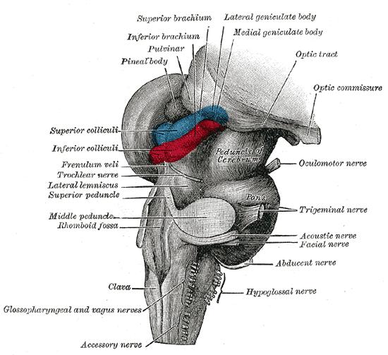
1. Noyau géniculé latéral : le noyau géniculé latéral est un noyau du thalamus postérieur. Il est situé sur la surface ventrale du thalamus, sur la face externe des pieds cérébraux et sur la partie supérieure du corps géniculé médian.
2.Diencephalon: Diencephalonislocatedbetweenthemidbrainandthetelencephalon.Thediencephalonandthetelencephalonbothderivefromtheforebrainalarintheearlyembryonicstage.Thediencephalonislocatedinthebackcenteroftheforebrain, andthetelencephalondevelopsintotheleftandrightcerebralhemispheres.Duetothehighexpansionofthetelencephalon, thediencephalonissurroundedbytheleftandrightcerebralhemispheresexceptforthepartthatbelongstothehypothalamusontheventralside, whichisexposedonthesurfaceofthebrain.Inthemid-sagittalsectionofthebrain, thelinefromtheposteriorcommissuretotheposterioredgeofthepapillarybodyrepresentsthejunctionbetweenthediencephalonandthemidbrain, andthelinefromtheinterventricularforamentotheopticchiasmrepresentsthejunctionbetweenthediencephalonandthetelencephalon.Theventricularcavityofthediencephaloniscalledthethirdventricle.

Theinnersideofthediencephalononbothsidesformsthesidewallofthethirdventricle.Atthejunctionofthemedialanddorsalside, thereisaraisedfiberbundle-thalamicmedullastriatum, whichisattachedtothethirdventriclechoroidtissue.Themedullaryveinsofthethalamusconnectthereintriangleback, andthereisareincommissureconnectionbetweentheleftandrightreintriangles, andthereisapinealglandbehindthiscommissure.Approximatelyinthemiddleofthethirdventriclewall, thereisanadhesive (orcentralmass) betweenthethalamusconnectingtheleftandrightventricularwalls.Ithasahypothalamicsulcusonitsventralside, fromthemidbrainaqueducttotheinterventricularforamen.Thestructurebelongingtothehypothalamussurroundsthefloorofthethirdventricle, andfromfronttobacktherearetheopticchiasm, thefunnel, thegraynodulesandthenipplebodyconnectingtheendplates.Onthebackofthediencephalon, bothsidesofthethirdventricleareheldbyovalgraymattermassesbelongingtothedorsalthalamus, withaprotrudinganteriorthalamicnoduleatthefrontendandanenlargedoccipitalattheback.Thereisthe caudatenucleusbelongingtothetelencephalonontheoutersideoftheback, andthestriaterminalisbetweenitandthediencephalon.Onthelowerandoutersideofthepillow, therearemedialgeniculatebodyandlateralgeniculatebody.Thelateralsurfaceofthediencephalonisfusedwiththeinternalcapsuleofthetelencephalon.Theventralsurfaceofthediencephalonisthepartexposedontheoutersurfaceofthebrain, withtheopticchiasmandoptictractbeforeit.Thefunnel, pituitaryandgraynodulesareinthemiddle, andthepapillarybodiesarepairedandlocatedbehindthegraynodules.
Anatomie
Lateralgeniculatebody: Thelateralgeniculatebodyislocatedunderthethalamusocciput, andthemedialgeniculatebodyisontheoutside, oneontheleftandoneontheleftandoneontheleftandoneontheleftandright.Thegroup, calledthelateralgeniculatenucleus, isthesubcorticalcenterofvision, connectedtothesuperiorcolliculusofthemidbrainquadrantviathesuperiorcolliculusarm.Thelateralgeniculatebodyisapairofcranialnervesdirectlyconnectedwiththediencephalon, thatis, theterminalnucleusoftheopticnerve (Ⅱ) .Thefibersemittedbythenucleusformopticradiationandreachthevisualcenterofthecerebralcortex.Itisthelastrelaynucleusonthevisualconductionpathway ..
Lastructuremorphologiqueetlacompositionducorpsgéniculélatéral
Thelateralgeniculatebody: Thelateralgeniculatebodyisalsocalledtheexternalgeniculatebody.Thesmallellipticalbumpslocatedontheoutsideofthefeetofthebrainandundertheoccipitaloftheopticthalamusbelongtothediencephalonandarethefirst-levelvisualcenterofthevisualanalyzer.Theopticfibersoftheoptictractstopattheganglioncellsofthelateralgeniculatebodyandexchangeneuronstoformopticradiation, whichallprojecttothestriateareaof thevisualcenteronthesamesidetoproducevision.Theopticalfibersinthelateralgeniculatebodyalsohaveacertainarrangement.Theperipheraluncrossedfibersfromtheipsilateralretinaandthecontralateralcrossedfibersterminateontheventralsideofthelateralgeniculatebody.Theupperhalfofthefiberislocatedontheventralinnersideandthelowerhalf.Thefibersarelocatedontheventralside.Themacularfiberisfinallydorsal.Theupperquadrantmacularfiberislocatedinsidethewedge-shapedarea, andthelowerquadrantmacularfiberislocatedoutsidethewedge-shapedarea.VonMonakowdividesthelateralgeniculatebodyintothreeparts, à savoir: ①thelowerp artdelapartieantérieure,quiestconnectéeaufaisceauoptique;②lapartieporte;③lapartielatérale,quiestlapureducorpsgéniculélatéral.
Les caractéristiques et l'importance clinique de l'apport sanguin du corps géniculé latéral
Thesmallarteriesofthelateralgeniculatebodyaremulti-sourceandmulti-vesselbloodsupply, 80% ofwhicharemorethandualsources, smallarteriesAsmanyas3-6, thesesmallarteriesfromdifferentsourcesareanastomosedwitheachotheronthesurfaceofthelateralgeniculatebody.Webelievethatevenifthereare1-2tertiarybloodsupplyarteriesblockedornarrowed, itwillnotaffectthenutritionofthelateralgeniculatebodyandvisualfielddefects.Thepossibilityisunlikely.However, 20% ofthesinglesourcearterialblooddonorshaveimportantsignificanceintheetiologyofvisualfielddefects.Forexample, thesourcearterialatherosclerosiscanblocktheartery, whichcancausethelateralgeniculatebodytobecomeischemicandlosetheconductionfunction.
